GeoMx and mycosis fungoides
Project on lymphoma
Mycosis fungoides, or cutaneous T-cell lymphoma, is a cancer in which deranged T lymphocytes start proliferating in the skin. The client carried out the wet part of the study using GeoMx technology and ordered the analysis from us at BostonGene. I led the project, worked out the annotations and the nuances of the technology and communicated with the client, while Lesha did the quality control and analysis.
GeoMx technology and files
Original article: Multiplex digital spatial profiling of proteins and RNA in fixed tissue, 2020
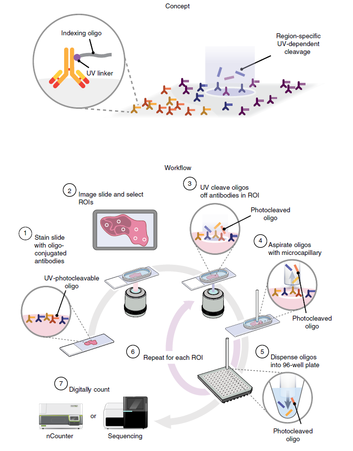
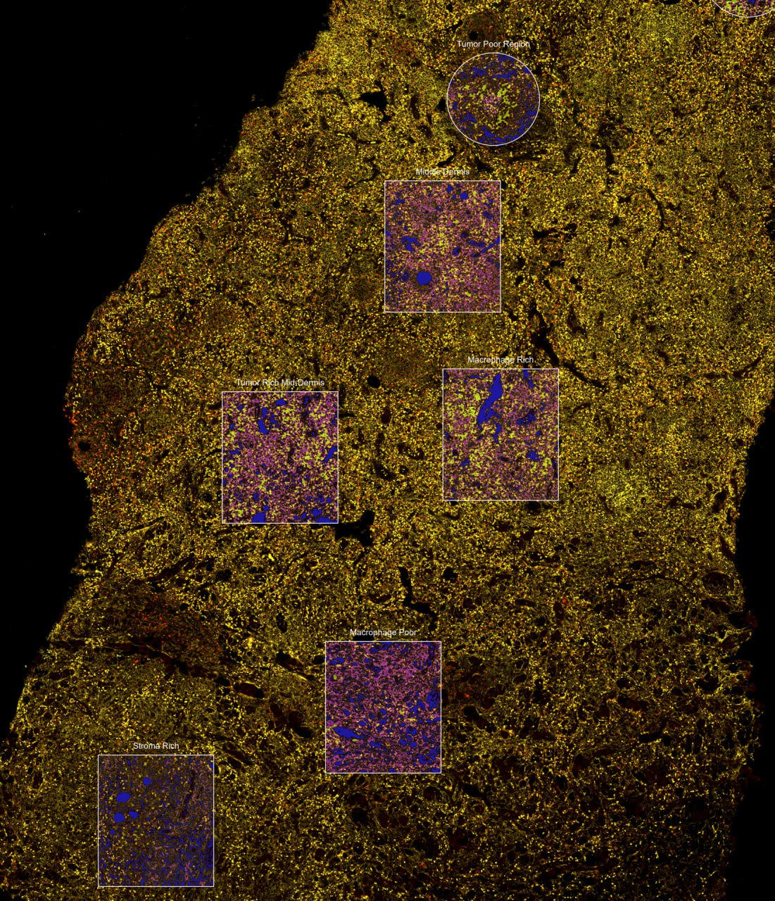
The essence of GeoMx technology:
- There is a pre-prepared set of RNA probes for thousands of genes. These probes are connected by a UV linker to a barcode oligonucleotide. One gene we care about = one barcode.
- The biopsy is prepared, placed on a slide, treated with RNA probes, and hybridization takes place.
- Then the slide is placed into a machine with a camera, and on the microphoto shown on the machine’s screen, the researcher marks the region of interest.
- Then the machine blasts this outlined region with UV light, the UV linker falls apart, and the machine sucks up the barcodes.
- The barcodes are sequenced, even on a regular sequencer, and reads are produced.
- The reads are matched against a pre-prepared barcode–gene panel. The number of reads from each barcode is basically transformed into the signal intensity of a particular RNA.
Therefore when we received raw sequencing files it became clear that this was not what we needed. We were sent oligo‑DNA barcodes that by themselves carry no information about genes and need to be matched to pairs of barcode‑gene via a special pipeline. The output files are Digital Count Conversion (DCC).
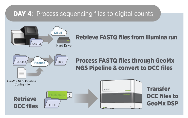

You can process fastq files yourself via Illumina’s BaseSpace GeoMx NGS Pipeline is distributed as a pipeline on BaseSpace. It seems this doesn’t require special access, but it costs money and is not trivial at all. So I decided to write to the collaborator and ask if they already had DCC files. The DCC files were received the next day. Sometimes it’s easiest to simply ask.
Annotation
To ask the right questions about the cohort and then answer them you need to understand what you’re dealing with in the first place. In theory each patient may have several slides, and each slide may contain several ROIs. And patients should have different properties, something to compare with something.
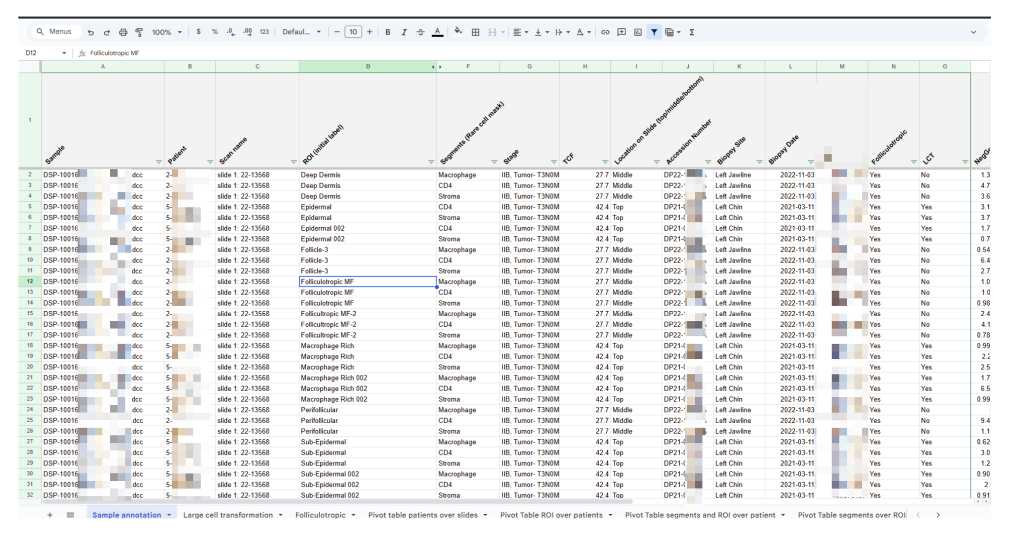
After studying the table it became clear which dimensions are important:
- Patient — 1st dimension
- Diagnosis and Large‑cell transformation — 2nd dimension
- Segments — 3rd dimension
- ROI – region of interest or mask — 4th dimension
Now this needs to be visualized.
1st dimension. We depicted the patient as a little man —
2nd dimension. We group patients by clinical parameters
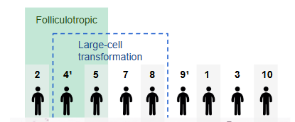
3rd dimension. In the annotation there are names “Epidermal”, “Sub‑epidermal”, “Dermis”. Therefore I decided that investigators took segments according to the principle “look at the skin at different depths”. I chose a picture of “skin in cross‑section” with the upper, middle and deep structures and hair follicles.
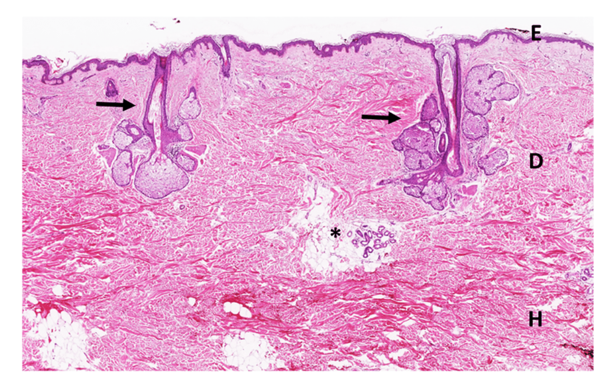
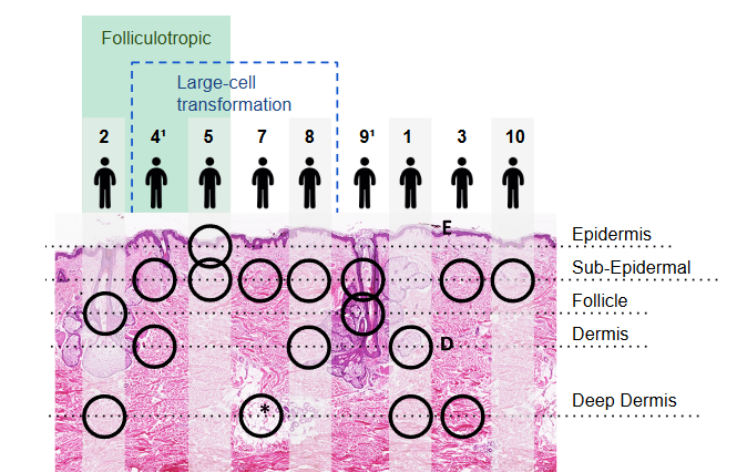
Now the data structure is clear. For example, only patient 5 has an Epidermal segment, or follicles appear only in two patients but they have different diagnoses.
In the course of the exercise it turned out that segment names sometimes designated localization – “Epidermal”, and sometimes a property – “Macrotumor poor”. To clarify the annotation I prepared a table where categories for ROI were partially unified. We asked collaborators to fill in the orange cells and prepared a slide with an explanation of what was unclear.
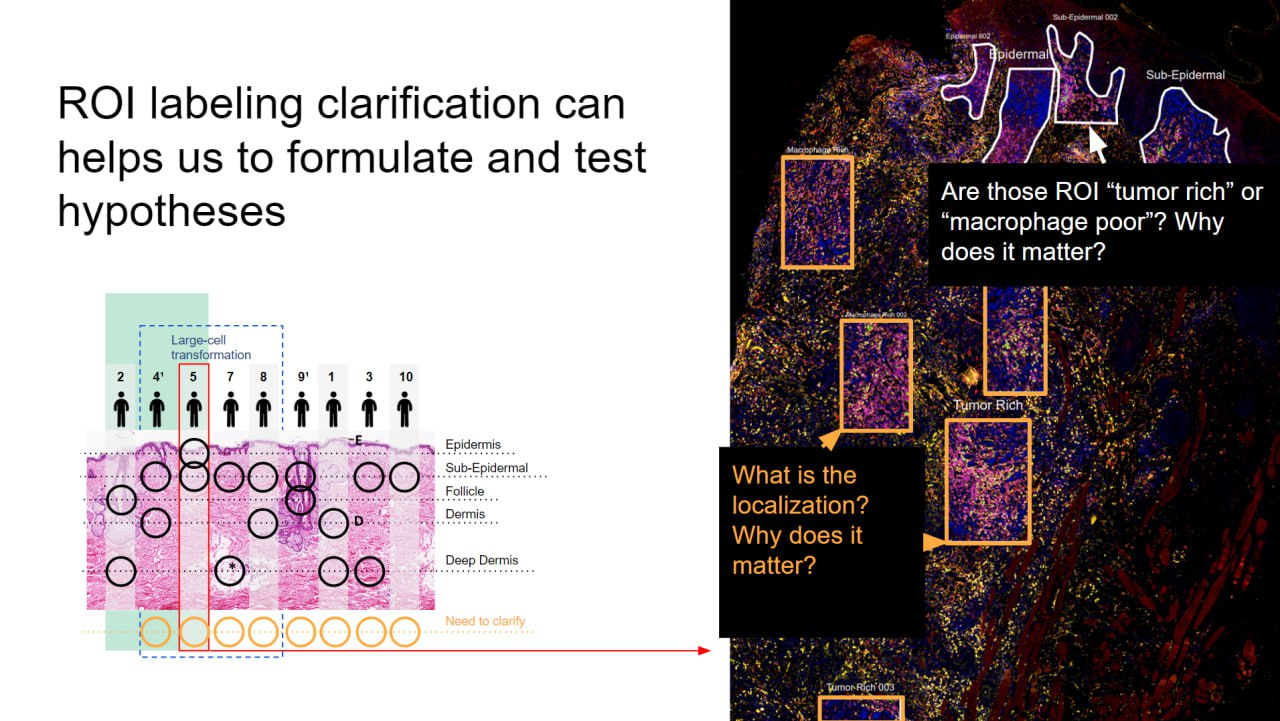
We wrote a letter to the client, explained the situation and asked them to clarify the annotation.
Analysis and results
Lesha immersed himself in the technology, selected tools for work and began the analysis: Background modeling, Quality Control and Normalization for GeoMx RNA data with GeoDiff. Having understood the technology, the necessary files and the structure of the cohort, the rest went without problems.
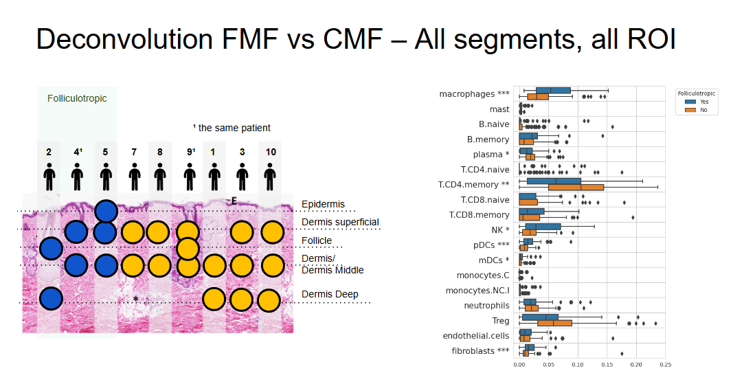
We found that FMF has an overall inflammatory microenvironment with a predominance of components of innate immunity – T1 and M2 macrophages, NK cells and dendritic cells. And CMF is less inflamed, with a predominance of T‑helper and plasma cells.
The client looked satisfied at the meeting, but then refused to pay us, and we forbade them to publish a paper with our analysis. We never learned the real reason. It happens.